Samples submitted to the UNL Plant and Pest Diagnostic Clinic and reports from clientele indicate that additional leaf diseases have begun to develop in parts of Nebraska following recent warm, wet, and/or humid weather conditions.
There are many questions right now on differentiating leaf diseases. Very early symptoms of many diseases look similar. For example, small yellow flecks may be indicative of several diseases and it can be hard to differentiate them without the presence of more mature lesions (or use of a microscope to examine spores and bacterial streaming). This article lists several leaf diseases that have been confirmed or suspected in Nebraska in recent days.
Risk Factors
Risk factors for leaf disease in corn include:
- susceptible hybrid,
- continuous corn,
- history of severe disease,
- minimum or no-till, and
- recent or forecasted wet weather or high relative humidity.

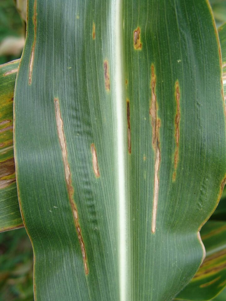
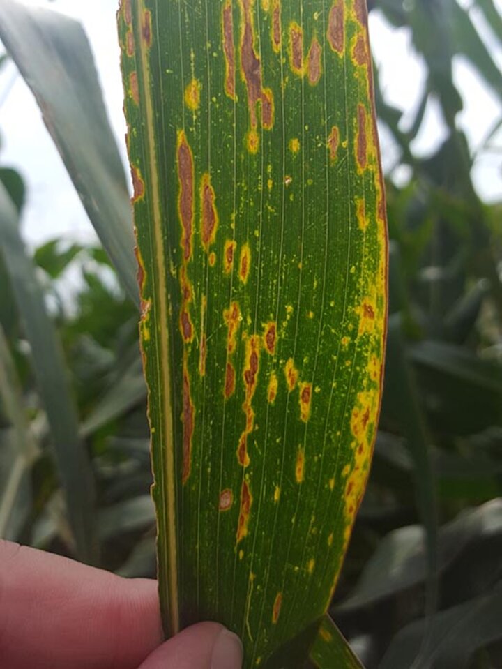
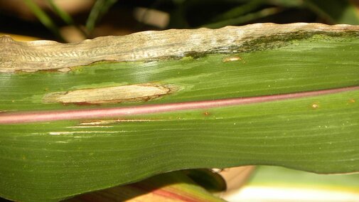
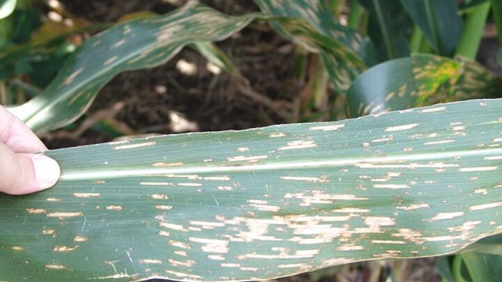
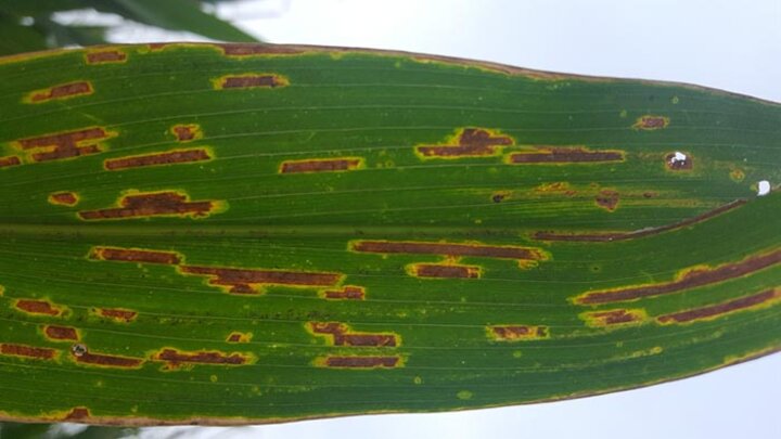
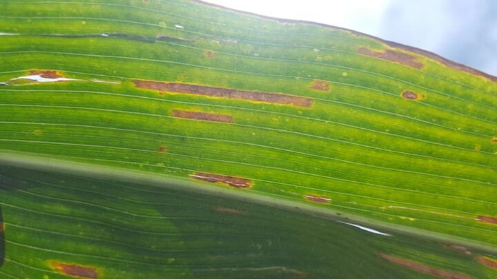
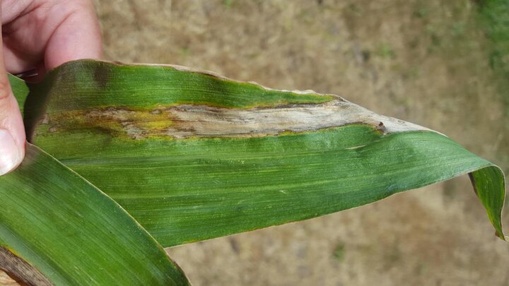
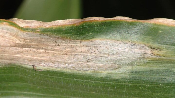
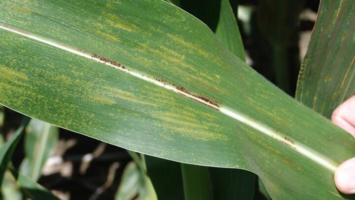
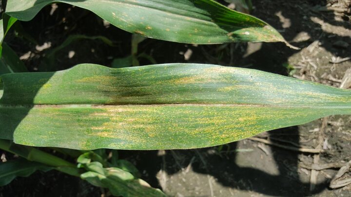
Bacterial Leaf Streak. Bacterial leaf streak (Figures 1-4) is common in many parts of Nebraska now. Lesions usually begin developing on lower leaves of the plant. They are yellow to tan streaks between the veins and often have a yellow hue, especially when backlit. Lesions are most often confused with those of the fungal disease gray leaf spot. Bacterial leaf streak lesion margins are usually more irregular or wavy in contrast to the smooth, straight lesions of gray leaf spot.
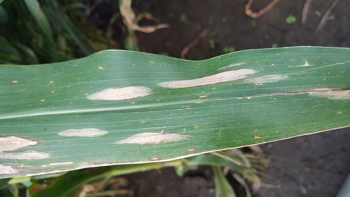
Gray Leaf Spot. The fungal disease gray leaf spot is a common disease during most years across much of Nebraska and has already begun to develop in some parts of the state following wet/humid and warm weather conditions. The gray to tan lesions develop between the veins and are distinctly rectangular with smooth, linear margins along the leaf veins. Lesions are slow to develop, needing 14-21 days, and begin in the lower leaves. Monitor high risk fields closely and consider the risk factors and weather conditions before making fungicide application decisions.
Goss’s Bacterial Wilt and Blight. The bacteria causing Goss’s “wilt” often use wounds caused by hail, wind, etc. to infect plants. Lesions can be large and watersoaked with very irregular shapes. The edges of lesions often may have dark spots or “freckles” that are diagnostic for the disease, as well as shiny bacterial exudate on either the upper or lower leaf surface.
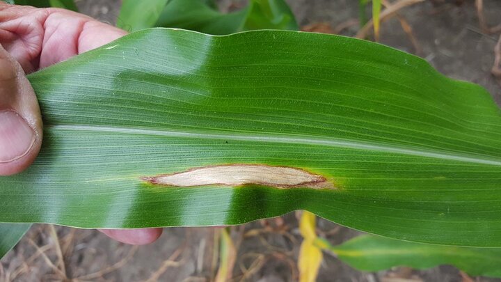
Northern Corn Leaf Blight. Northern corn leaf blight is caused by another fungus that overwinters in infested crop debris. The tan lesions can be small to very large and elliptical in shape, usually with smooth round ends. Mature lesions in humid conditions may appear dusty in the middle as the fungus produces spores that are spread to other leaves.
Physoderma brown spot. Physoderma brown spot is caused by a fungal-like organism that overwinters in crop debris. The organism produces specialized swimming spores that use water to infect the plant, thus wet conditions are necessary for infection. Infection often occurs in the whorl. As leaves emerge through the whorl during alternating wet/dry cycles, leaves can be infected, causing bands of lesions across the leaves. The lesions vary in appearance depending on where they occur. Lesions on the leaf blade are tiny and yellow to tan in color and sometimes mistaken for southern rust. However, the lesions produced in the midrib often appear much larger and dark brown to back in color. The presence of these two types of lesions in bands on the same leaf is a helpful clue when identifying the disease. Spores also may be washed down the leaf sheath and accumulate at the nodes where they may also infect. Nodal infections can weaken stalks, making them more brittle and vulnerable to lodging.
Need help? Instructions and a form for submitting samples can be found on the UNL Plant and Pest Diagnostic Clinic page.